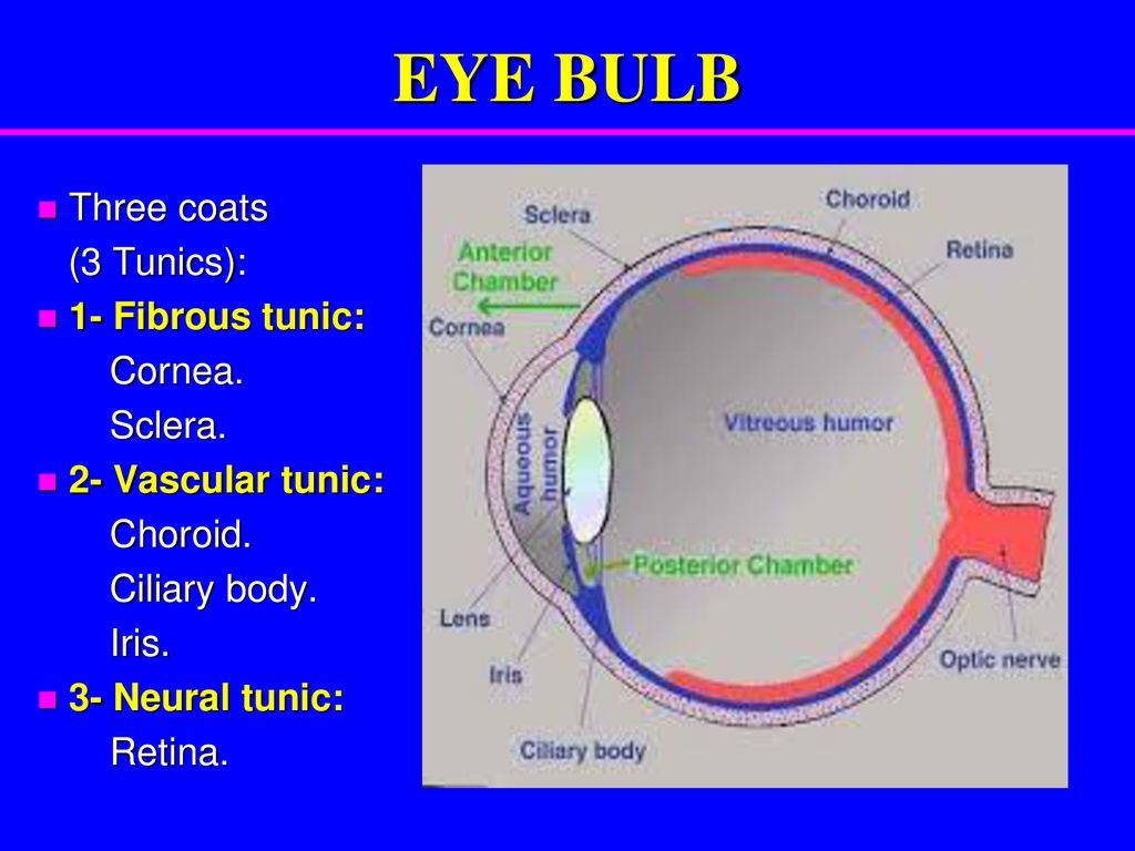
Tunics Of The Eyeball
The sclera is opaque and constitutes the posterior five sixths of the tunic. Play this quiz called tunics of the eyeball and show off your skills.
Mrs Amany Ahmed Niazy Opto 435 Lecture 2 Eyeball The Eyeball
Internal nervous tunic retina.

Tunics of the eyeball. From outermost to innermost these are the corneoscleral tunic the uveal tunic and the retinal tunic. The sclera and cornea form the fibrous tunic of the bulb of the eye. The tunics of the eyeball the organ of vision of vertebrate animals is enclosed in a firm case of fibrous tissues called the sclerotic coat which is continuous with the sheath of the optic nerve.
And 3 a nervous tunic the retina. 2 a vascular pigmented tunic comprising from behind forward the choroid ciliary body and iris. Anatomically the eyeball can be divided into three parts the fibrous vascular and inner layers in this article we shall consider the anatomy of the eyeball in detail and its clinical correlations.
It lies in a bony cavity within the facial skeleton known as the bony orbit. The vascular tunic also known as the tunica vasculosa oculi or the uvea is the middle vascularized layer which. 869 consisting of the sclera behind and the cornea in front.
It is seen between the eyelids under the transparent conjunctiva and known as the white of the eye. From without inward the three tunics are. Tunics of eye external fibrous tunic.
The sclera and cornea form the fibrous tunic of the bulb of the eye. The sclera is opaque and constitutes the posterior five sixths of the tunic. This is a quiz called tunics of the eyeball and was created by member shandelly22 english en.
Fibrous tunic of eyeball. Horizontal section of the eyeball. 1 a fibrous tunic fig.
Three layers the fibrous tunic also known as the tunica fibrosa oculi is the outer layer of the eyeball consisting of the cornea. The eyeball is a bilateral and spherical organ which houses the structures responsible for vision. The three tunics of the eye the wall of the eyeball is made up of three coats or tunics one inside the other.
869 horizontal section of the eyeball. The middle tunic frequently termed the uvea comprises the choroid the ciliary body and the. The nervous tunic also known as the tunica.
Cornea labeled at top sclera labeled at center right details. The corneoscleral tunic s functions are chiefly mechanical and optical. Tunica fibrosa bulbi tunica fibrosa oculi.
The cornea is the anterior transparent part of the eye and it forms about one sixth of the. The tunics of the eye. The cornea is transparent and forms the anterior sixth.
The cornea is transparent and forms the anterior sixth.
Major Ocular Structures Laramy K Independent Optical Lab
Ocular Anatomy Flashcards Easy Notecards
Eye Anatomy Flashcards Quizlet
The Eye Neuro Microanatomy The Eye Flashcards Memorang
Detailed Structures Of The Eyeball Video Lesson Transcript
Senses Part 2 Tunics Of The Eye Fibrous Tunics Sclera
Nervous Tunic Definition Of Nervous Tunic By Medical Dictionary
Http Semmelweis Hu Anatomia Files 2018 09 Tzs Fibrous And Vascular Coats Of The Eyeball 2018 Pdf
P Essentials Of Anatomy Seeley Stephens Tate Special Senses Part
The Tunics Of The Eye Human Anatomy
The Tunics Of The Eye Human Anatomy
The Eye Overview And Terminology
Anatomy Of The Eye Flashcards Quizlet
Anatomy Of The Eye Veterinary Medicine 2 Year With Several At
Histology Of The Eye Ppt Download
Ppt The Special Senses Powerpoint Presentation Free Download
The Tunics Of The Eye Human Anatomy
Anatomy Of The Internal Eye Sagittal Views Depict A The Three
Https Encrypted Tbn0 Gstatic Com Images Q Tbn 3aand9gcqwinimao3drgpsiqsjgtlde8umacnvaqgsgds0bhc3wijhn9xf Usqp Cau
Eye Anatomy And Vision Course Hero
The Tunics Of The Eye Human Anatomy
The Tunics Of The Eye Human Anatomy
There Are Three Layers Or Tunics Of The Eyeball The Fibrous
Eyes Eyes And Their Functions Structure Of The Eyes
The Eye Ear Special Sense Organs Junqueira S Basic Histology
Anatomy Of The Eyeball And 3 Tunics Youtube
Posting Komentar
Posting Komentar