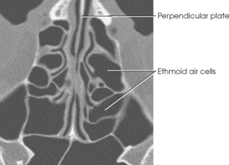
Perpendicular Plate Of Ethmoid Bone Ct
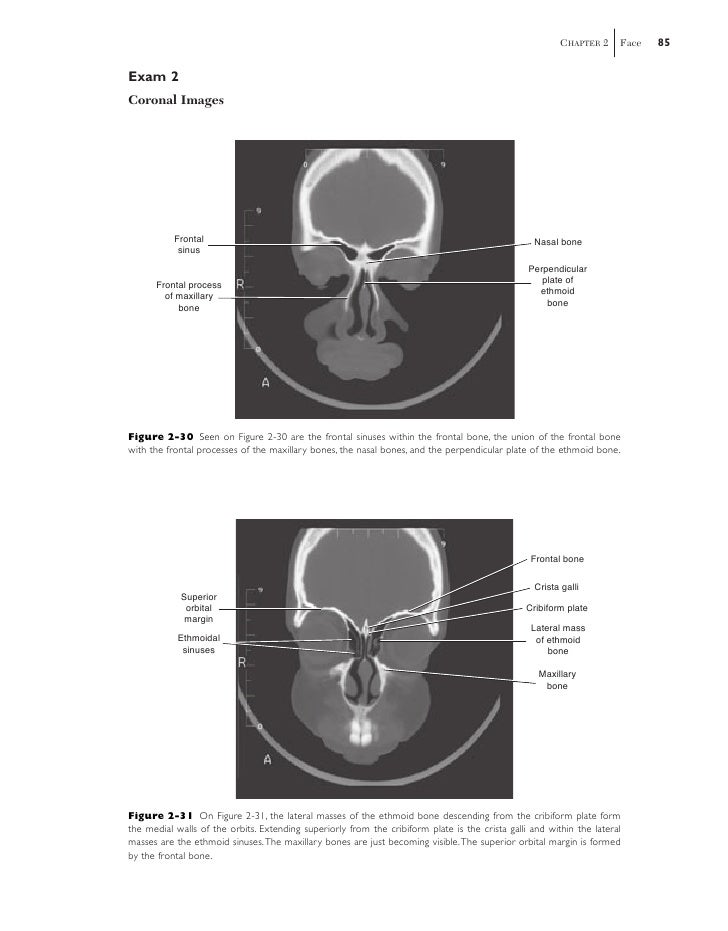
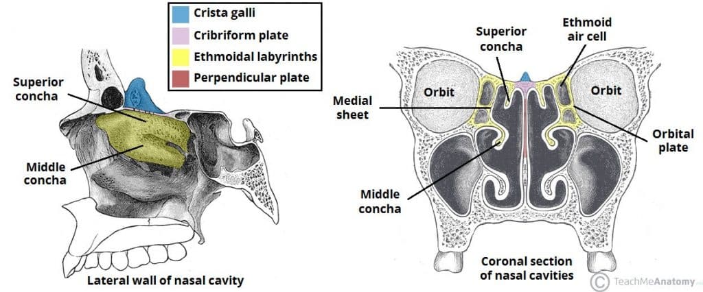
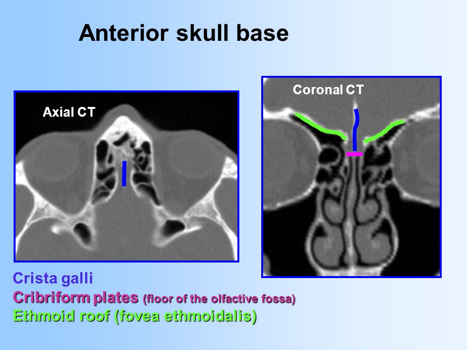
In the center of the ethmoid bone between the ethmoidal labyrinth is the perpendicular plate which forms the upper part of the bony nasal septum. The olfactory bulbs of the olfactory nerve lie on either side of the crista galli on top of the cribriform plate.
Ethmoid Bone Anatomy Borders And Development Kenhub
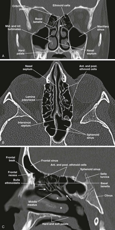
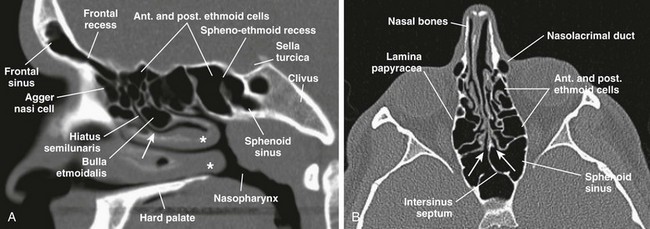
Ethmoid bulla bulla ethmoidalis medial rectus muscle.
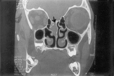
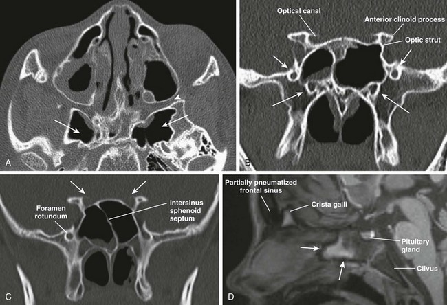
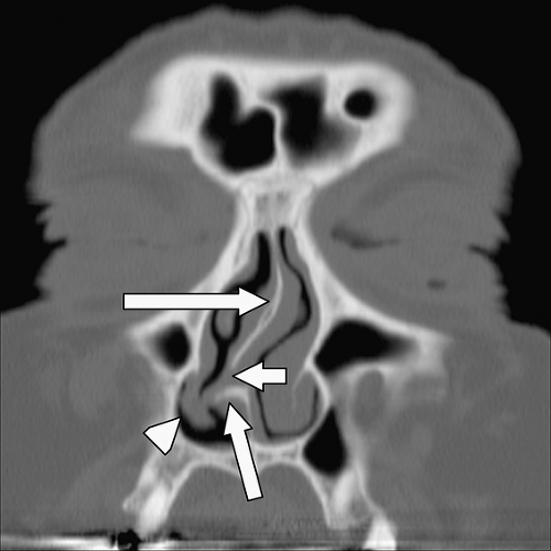
Perpendicular plate of ethmoid bone ct. Six cases 6 32 18 were found to have pneumatization of perpendicular plate of the ethmoid bone with 2 located in the anterior portion frontal septal pneumatization and 4 located in the posterior portion spheno septal pneumatization. Sella turcica pituitary fossa temporalis muscle. The horizontal plate forms the posterior portion of the hard palate of the oral cavity and is directly inferior to the nasal cavity.
The ct data from 32 patients with septal deviation were reviewed and an unusual case of perpendicular plate mucocele was reported. This medial area contains the greater palatine. These are large masses located at either side of the perpendicular plate which contain the ethmoidal air cells sinuses.
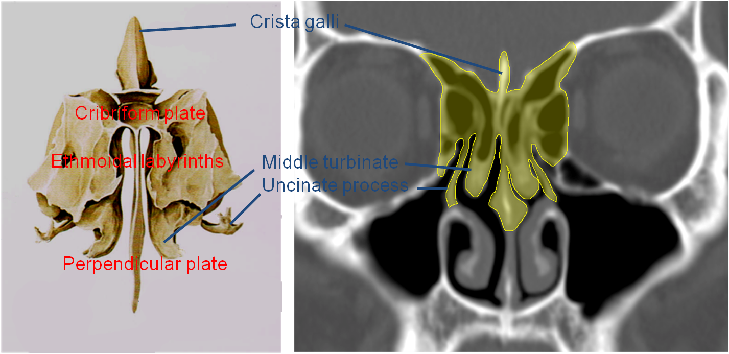
Crest of the rooster is the upper part of the perpendicular plate of the ethmoid bone which rises above the cribriform plate. The ethmoid bone consists of four parts. Ethmoid bulla bulla ethmoidalis medial rectus muscle.
The labyrinths are first developed ossific granules making their appearance in the region of the lamina papyracea between the fourth and fifth months of fetal life and extending into the conchæ. The palatine bone consists of a horizontal and perpendicular plate and the pyramidal process. The lower part of your nasal septum is formed by the vomer bone and the palatine bone.
One for the perpendicular plate and one for each labyrinth. The posterior nasal spine sits at the back of the horizontal plate where the two opposing palatine bones articulate. Perpendicular plate of the ethmoid bone.
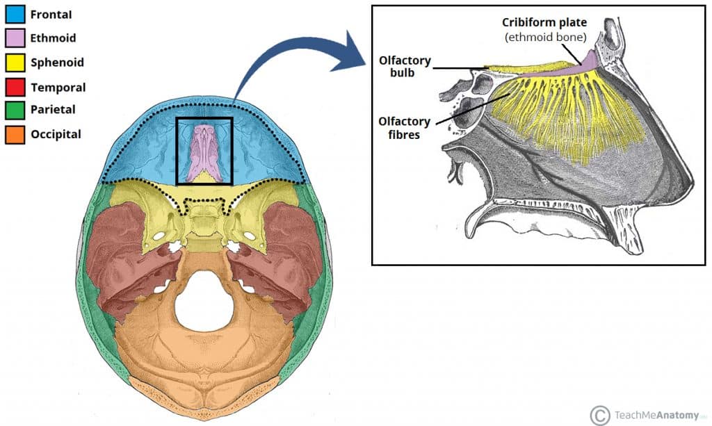
It contributes to the anterior cranial fossa. Sella turcica pituitary fossa temporalis muscle. The ethmoid bone is a single midline facial bone that separates the nasal cavity from the brain and is located at the roof of the nose and between the orbits it is a cubical shape and is relatively lightweight because of its spongy construction.
The anterior border articulates with the spine of the frontal bone and the crest of the nasal bones. The falx cerebri fold of the dura mater attaches to the crista galli. The ethmoid is ossified in the cartilage of the nasal capsule by three centers.
It forms the superior two thirds of the nasal septum. It is generally deflected a little to one or other side. Perpendicular plate of the ethmoid bone.
The crista galli latin. The perpendicular plate of the ethmoid bone vertical plate is a thin flattened lamina polygonal in form which descends from the under surface of the cribriform plate and assists in forming the septum of the nose. Lastly the ethmoid bone contains two ethmoidal labyrinths.
Another projection of bone descends from the cribriform plate the perpendicular plate.


Lecture 1 Terminology Skull Orbit Head And Neck Flashcards
Medial Wall Of The Nasal Cavity Anatomy And Structure Kenhub


Ethmoid Bone Anatomy Borders And Development Kenhub


Http Pdf Posterng Netkey At Download Index Php Module Get Pdf By Id Poster Id 115345


Https Encrypted Tbn0 Gstatic Com Images Q Tbn 3aand9gct Q33ytul Dwpz5 5zrniwpiijwuigibjhdhabpmqdnyjqbmp Usqp Cau


Posting Komentar
Posting Komentar